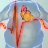
True Aneurysm of Extracranial Internal Carotid Artery in a 10-year-old
- Çocuklarda oldukça nadir görülen ekstrakraniyal karotid arter gerçek anevrizmaları, boyun kitlelerinin ayırıcı tanısında hayati bir risk faktörü olarak değerlendirilmelidir.
- 10 yaşındaki bir hastada saptanan 25x50 mm boyutundaki anevrizma, cerrahi müdahale ile rezeke edilerek sentetik ePTFE greft interpozisyonu yöntemiyle başarıyla tedavi edilmiştir.
- Tedavi edilmeyen karotid anevrizmaları %50'ye varan felç ve yüksek mortalite riski taşıdığından, cerrahi sonrası düzenli Doppler ultrasonografi takibi büyük önem arz etmektedir.

İçerik yapay zeka ile optimize edilmiştir
Giriş: Çocuklarda Ekstrakraniyal Karotid Arter Anevrizmaları
Ekstrakraniyal karotid arterlerde görülen aterosklerotik tıkayıcı hastalıklar yetişkinlerde yaygın olsa da, bu damarların gerçek anevrizmaları oldukça nadir görülen patolojilerdir. Özellikle herhangi bir eşlik eden hastalık öyküsü bulunmayan pediatrik popülasyonda gerçek anevrizma vakalarına rastlanması istisnai bir durumdur. Bu nadir duruma rağmen, boyun kitlelerinin ayırıcı tanısında ekstrakraniyal karotid anevrizmaları mutlaka göz önünde bulundurulmalıdır.
Olgu Sunumu: 10 Yaşındaki Hastada İnternal Karotid Arter Anevrizması
Dr. Siyami Ersek Göğüs Kalp ve Damar Cerrahisi Merkezi'ne başvuran 10 yaşındaki erkek hastada, boynun sağ tarafında pulsatil (vuru veren) bir kitle tespit edilmiştir. Hastanın bir yıldır devam eden boyun ağrısı ve baş ağrısı şikayetleri mevcuttur. Geçmişinde travma, vaskülit veya kronik bir hastalık öyküsü bulunmayan hastanın, sadece 2004 yılında sorunsuz bir tonsillektomi operasyonu geçirdiği öğrenilmiştir.
Tanı ve Klinik Bulgular
Yapılan fizik muayenede, boyun sağ lateral bölgesindeki kitlenin pulsatil karakterde olduğu ve üzerinde üfürüm (bruit) duyulduğu saptanmıştır. Tanısal süreci netleştirmek adına uygulanan tetkiklerde şu bulgulara ulaşılmıştır:
- Servikal Doppler ve Kontrastlı BT: Sağ internal karotid arterin bifurkasyon distalinde 25x50 mm boyutlarında gerçek anevrizma.
- Klinik Belirtiler: Hassas kitle, kronik boyun ağrısı ve tekrarlayan baş ağrıları.
Cerrahi Teknik ve Uygulama
Hasta operasyon hazırlığı kapsamında hastaneye yatırılarak 24 saatlik sodyum heparin infüzyonuna alınmıştır. Genel anestezi altında, standart anterior sternokleidomastoid insizyonu ile cerrahiye başlanmıştır. Ameliyat sırasında uygulanan teknik detaylar aşağıda maddelenmiştir:
- Uzun disseksiyon süresi öngörüldüğü için arteriyotomi sonrası şant yerleştirmesi yapılmıştır.
- Anevrizmatik segmentin rezeksiyonu titizlikle gerçekleştirilmiştir.
- Safen ven çapının yetersiz olması (5 mm altı) nedeniyle, 5 mm ePTFE prostetik greft interpozisyonu tercih edilmiştir.
- Şantın greft içinden geçirilerek proksimal ve distal uçlara yerleştirildiği pragmatik bir teknik kullanılarak cerrahi saha hakimiyeti artırılmıştır.
| Parametre | Detaylar |
|---|---|
| Hasta Yaşı/Cinsiyet | 10 Yaş / Erkek |
| Anevrizma Boyutu | 25 x 50 mm |
| Kullanılan Greft | 5-mm ePTFE |
| Hastanede Kalış Süresi | 3 Gün |
Tartışma ve Literatür Değerlendirmesi
Karotid arterlerin gerçek anevrizmaları, aterosklerotik lezyonlara kıyasla hem daha nadirdir hem de yönetimi daha karmaşıktır. İnternal karotid arter, ana karotid bifurkasyonundan sonra anevrizmaların en sık görüldüğü ikinci lokasyondur. Yetişkinlerde etyoloji genellikle ateroskleroz, travma, disseksiyon veya enfeksiyon kaynaklıyken, çocuklarda bu durumun örneği çok azdır.
Ayırıcı Tanının Önemi ve Riskler
Bu patolojinin hızlı ve doğru teşhis edilmesi hayati önem taşır. Yanlışlıkla parafaringeal apse tanısı konulması veya teşhiste geç kalınması; yaşamı tehdit eden kanamalara veya kalıcı felçlere (stroke) yol açabilir. Tedavi edilmeyen vakalarda felç oranı %50 seviyelerine çıkabilmekte ve yüksek mortalite riskine neden olmaktadır.
Sonuç
İnternal karotid arter anevrizmaları, cerrahi yöntemlerle başarılı bir şekilde yönetilebilir. Otojen ven greftleri ilk tercih olsa da, gerekli durumlarda sentetik greftler güvenle kullanılabilir. Bu vakada, hastanın büyüme potansiyeli dikkate alınarak hafif uzun bir greft yerleştirilmiştir. Ameliyat sonrası dönemi sorunsuz atlatan hasta, 3. günde taburcu edilmiştir. Bu tür vakalarda ilk yıl boyunca her üç ayda bir Doppler ultrasonografi ile takip, uzun dönemli başarı için kritiktir.