The Reliability of the Ultrasonic Bone Scalpel in Cervical Spondylotic Myelopathy: A Comparative Study of 46 Patients

İçerik yapay zeka ile optimize edilmiştir
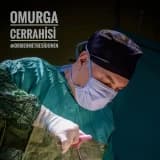
Servikal Spondilotik Miyelopati (CSM) ve Laminektomi Teknikleri
Servikal spondilotik miyelopati (CSM), spinal kordda meydana gelen hasara bağlı olarak gelişen motor ve duyusal fonksiyon kaybıdır. Bu durum genellikle dejeneratif patolojilerle ilişkili akut veya kronik bir süreç olarak ortaya çıkar. CSM tedavisinde en sık tercih edilen cerrahi yöntemlerden biri posterior laminektomi işlemidir.
Geleneksel laminektomi prosedürlerinde genellikle yüksek hızlı drill (HSD) ve Kerrison rongeur kullanılmaktadır. Ancak bu araçlar, özellikle dar spinal kanallarda dura yaralanması, termal hasar ve mekanik nöral yaralanma gibi riskler taşımaktadır. Son yıllarda geliştirilen ultrasonik kemik bistürisi (UBS), kemik dokusunu ses dalgalarıyla keserek yumuşak doku hasarını minimize eden daha güvenli bir alternatif sunmaktadır.
Araştırma Yöntemi ve Hasta Grupları
Bu çalışma, 2010-2014 yılları arasında CSM tanısıyla ameliyat edilen 46 hasta üzerinde retrospektif olarak gerçekleştirilmiştir. Hastalar kullanılan cerrahi enstrümana göre iki eşit gruba ayrılmıştır:
- Grup I (UBS Grubu): 23 hastaya ultrasonik kemik bistürisi ile laminektomi uygulanmıştır.
- Grup II (HSD Grubu): 23 hastaya yüksek hızlı drill ile laminektomi uygulanmıştır.
Her iki grupta da demografik özellikler, laminektomi seviyeleri, operasyon süreleri, kan kaybı miktarları ve cerrahi komplikasyonlar titizlikle karşılaştırılmıştır. Klinik iyileşme oranları Japon Ortopedi Birliği (JOA) skorları ve Hirabayashi formülü kullanılarak analiz edilmiştir.
Bulgular: UBS ve HSD Performans Karşılaştırması
Yapılan incelemeler sonucunda, ultrasonik kemik bistürisi kullanımının cerrahi verimlilik ve hasta güvenliği açısından anlamlı avantajlar sağladığı saptanmıştır. Gruplar arasındaki temel veriler aşağıdaki tabloda özetlenmiştir:
| Parametre | Grup I (UBS Grubu) | Grup II (HSD Grubu) |
|---|---|---|
| Laminektomi Süresi (dk/seviye) | 2.2 ± 0.4 | 7.4 ± 2.6 |
| Ortalama Kan Kaybı (mL) | 180 (140-250) | 380 (280-450) |
| Hastanede Kalış Süresi (Gün) | 3.0 ± 0.0 | 3.7 ± 1.3 |
| Dura Yaralanması | Hiç yok | 3 hasta (%13.0) |
| C5 Radikülopatisi | 1 hasta | 1 hasta |
| İyileşme Oranı (JOA) | %47.6 | %48.8 |
Cerrahi Tekniklerin Uygulanışı
UBS tekniğinde, kemik kendi gücüyle kesilir ve iç korteks geçildikten sonra duranın yumuşaklığı hissedilerek işlem durdurulur. HSD tekniğinde ise lamina önce 2-3 mm'lik drill uçlarıyla inceltilir, ardından elmas uçlar ve cerrahi kancalar yardımıyla çıkarılır. Her iki yöntemde de tüm laminektomiler benzer cerrahi yetkinliğe sahip uzmanlarca uygulanmıştır.
Tartışma: Ultrasonik Kemik Bistürisinin Avantajları
UBS, kemiği saniyede 22.500 boylamsal titreşim yapan ses dalgalarıyla keser. Bu sistemin en büyük avantajı, dura ve ligamentum flavum gibi elastik yapıdaki yumuşak dokulara temas ettiğinde kesme işlemi yapmamasıdır. Bu özellik, iyatrojenik yaralanma riskini önemli ölçüde azaltır.
UBS kullanımının sağladığı başlıca faydalar şunlardır:
- Hızlı Dekompresyon: Seviye başına cerrahi süreyi yaklaşık %70 oranında kısaltır.
- Düşük Kan Kaybı: HSD grubuna göre kan kaybı yarı yarıya daha azdır.
- Termal Güvenlik: Sürekli su soğutma sistemi sayesinde kemik yüzeyinde yanık oluşumu engellenir.
- Minimal Kemik Kaybı: 1 mm'den daha ince kesi hattı oluşturarak, otojen greftleme için daha fazla kemik korunmasını sağlar.
Çalışmanın kısıtlılıkları arasında farklı drill uçlarının (elmas vs. kanallı) karşılaştırılmaması ve uzun dönem füzyon oranlarının takibi yer almaktadır. Ayrıca, UBS cihazının yüksek maliyeti rutin kullanım önündeki en önemli engeldir.
Sonuç
Servikal spondilotik miyelopati vakalarında posterior yaklaşım ile uygulanan laminektomide ultrasonik kemik bistürisi (UBS); güvenli, hızlı ve etkili bir dekompresyon sağlar. Dura yaralanması riskini minimize etmesi, operasyon süresini kısaltması ve kan kaybını azaltması sayesinde postoperatif morbidite oranlarını düşürür ve hastanede kalış süresini optimize eder. Maliyetlerin düşmesi durumunda, UBS'nin spinal cerrahide rutin bir enstrüman haline gelmesi öngörülmektedir.