Assessment of Differential Renal Function in Children with Hydronephrosis: Comparison of DMSA and MAG-3

İçerik yapay zeka ile optimize edilmiştir
Hidronefrozlu Çocuklarda Böbrek Fonksiyonlarının İzlenmesi
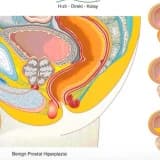
Pediatrik ürolojide sık karşılaşılan bir durum olan hidronefroz, idrar yollarındaki tıkanıklığa bağlı olarak gelişen ve ilerleyici böbrek hasarına yol açabilen bir tablodur. Bu hastaların tanısında, takibinde ve cerrahi müdahale kararının verilmesinde separe renal fonksiyonların (SRF) doğru hesaplanması kritik öneme sahiptir. Günümüzde bu amaçla en sık kullanılan nükleer tıp yöntemleri 99mTc-DMSA ve 99mTc-MAG-3 sintigrafileridir.
DMSA ve MAG-3 Sintigrafilerinin Temel Özellikleri
Böbrek fonksiyonlarını değerlendirmede kullanılan bu iki yöntemin kendine has avantajları ve uygulama alanları bulunmaktadır. DMSA sintigrafisi, böbrek kortikal yapısını ve kalıcı hasarları (skar) belirlemede altın standart olarak kabul edilirken; MAG-3 sintigrafisi, böbreklerin boşaltım dinamiklerini ve idrar akış paternini değerlendirmede üstündür.
Her iki yöntemin karşılaştırmalı özellikleri aşağıda sunulmuştur:
- DMSA (Dimerkaprosüksinik Asit): Kortikal bütünlüğü ve SRF'yi belirlemede referans yöntemdir. Enjeksiyondan 2-3 saat sonra görüntüleme yapılır.
- MAG-3 (Merkaptoasetilglisin-3): Renal klirens, tübüler fonksiyonlar ve boşaltım sistemi hakkında dinamik bilgi sağlar. Radyasyon dozu DMSA'ya göre yaklaşık 10 kat daha düşüktür.
Araştırma Yöntemi ve Hasta Grupları
Bu çalışmada, Ocak 2009 ile Ağustos 2013 tarihleri arasında izlenen 81 hidronefrozlu çocuk (toplam 102 böbrek ünitesi) retrospektif olarak incelenmiştir. Hastaların tanı anındaki yaşları, cinsiyetleri, renal pelvis AP çapları, parankim kalınlıkları ve her iki yöntemle elde edilen SRF değerleri kaydedilmiştir.
İstatistiksel analiz için hastalar şu kriterlere göre gruplandırılmıştır:
- SRF Değerleri: %45, %40 ve %10 kesme değerleri.
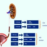
- AP Çapları: 5-10 mm, 10-20 mm, 20-30 mm ve 30-40 mm.
- Yaş: 24 ay altı ve 24 ay üstü.
Klinik Bulgular ve İstatistiksel Sonuçlar
Çalışma sonucunda, MAG-3 ve DMSA yöntemleri arasında SRF değerlerini belirleme açısından istatistiksel olarak anlamlı bir fark saptanmamıştır (p>0,05). Pearson korelasyon katsayısı r=0,926 olarak bulunmuş, bu da iki test arasında çok yüksek bir uyum olduğunu göstermiştir.
| Parametre | DMSA Sonuçları | MAG-3 Sonuçları | p Değeri |
|---|---|---|---|
| SRF ≤%45 | 20 Böbrek Ünitesi | 27 Böbrek Ünitesi | 0,065 |
| SRF ≤%40 | 14 Böbrek Ünitesi | 12 Böbrek Ünitesi | 0,687 |
| SRF ≤%10 | 2 Böbrek Ünitesi | 3 Böbrek Ünitesi | >0,05 |
Özellikle cerrahi karar aşamasında kritik olan %40 ve %10 kesme değerlerinde her iki testin sunduğu sonuçlar klinik olarak birbirini destekler niteliktedir. Ayrıca, hastanın yaşı veya hidronefrozun derecesi (AP çapı), testlerin birbirine olan üstünlüğünü değiştirmemektedir.
Sonuç: MAG-3 Tek Başına Yeterli mi?
Araştırma verileri, hidronefrozlu çocukların takibinde MAG-3 sintigrafisinin, separe renal fonksiyonları belirlemede DMSA kadar etkili olduğunu kanıtlamaktadır. MAG-3 kullanımı, çocukların gereksiz yere yüksek radyasyona maruz kalmasını engellemekte, zaman ve maliyet tasarrufu sağlamaktadır.
Önemli Çıkarımlar:
- MAG-3, hem fonksiyonel hem de dinamik veri sağladığı için çocuklarda ilk seçenek olarak değerlendirilebilir.
- DMSA sintigrafisi, yalnızca odak kortikal defektlerin şüpheli olduğu durumlarda ikincil test olarak saklanmalıdır.
- İki testin sonuçları, cerrahi endikasyon belirleme ve hasta takibi süreçlerinde klinik olarak uyumludur.